Best Radiographic Positions For Visualizing Anatomy
The quality of the radiographic image depends on several factors, including the position of the patient and the angle at which the x-ray beam is directed. To get the best possible image, it is important to position the patient correctly.
There are many different radiographic positions, and each one provides a different view of a particular area of the body. For example, the erect position is often used to image the chest or abdomen, while the supine position is better for imaging the pelvis or lower extremities.
In addition to positioning the patient correctly, it is also important to choose the appropriate x-ray projection. The most common projection is called the anterior-posterior (AP) projection, which creates a front-to-back view of the body. Other projections, such as lateral or oblique, can be used to get different views of the body depending on what Anatomy structures need to be seen.
Why Radiographic Positioning And Related Anatomy Is Necessary?
There are a few reasons why best radiographic positioning and related anatomy is necessary. First, it ensures that the radiograph is of high quality and will be diagnostically useful. Second, it minimizes the amount of radiation exposure to the patient. Third, it increases the chances that the radiograph will show the desired structures clearly and accurately. Finally, proper positioning often allows for better contrast between different tissue types, which can be critical for making an accurate diagnosis.
Our Top Picks For Best Radiographic Positioning And Related Anatomy


Best Radiographic Positioning And Related Anatomy Guidance
Textbook of Radiographic Positioning and Related Anatomy
Radiation therapy is the use of high-energy radiation to kill cancer cells. Radiation therapy can be delivered externally by a machine that focuses the radiation on the cancerous area, or internally by placing radioactive material directly into the cancerous tissue.
External radiation therapy is the most common type of radiation therapy used to treat cancer. The machine that delivers the radiation, called a linear accelerator, is large and complex. The linear accelerator uses electricity to produce high-energy X-rays that target the cancerous cells and kill them.
Internal radiation therapy involves placing radioactive material directly into the cancerous tissue. This type of radiation therapy is also called brachytherapy. The radioactive material is usually in the form of pellets, seeds, or wires that are placed into the cancerous tissue. The radioactive material emits radiation that kills the cancer cells.
Radiation therapy can be used to treat cancer at any stage. It is often used as part of a cancer treatment plan that includes surgery and chemotherapy. Radiation therapy can also be used to relieve symptoms caused by cancer.
The side effects of radiation therapy depend on the type and amount of radiation used, the part of the body that is treated, and the person’s overall health. Common side effects of radiation therapy
Common Questions on Textbook of Radiographic Positioning and Related Anatomy
• What is the CR for a pediatric chest x-ray?The CR for a pediatric chest x-ray is the anterior-posterior projection of the vertebral column. The CR should be perpendicular to the IR at the level of the xiphoid process.
• What are the standard SID for a chest x-ray?
The standard SIDs for a chest x-ray are 72 inches for an adult and 36 inches for a child.
• Which projection is used to visualize the heart?
The PA projection is used to visualize the heart. The CR should be perpendicular to the IR at the level of the xiphoid process and the SID should be 72 inches.
• How do you position a patient for a lumbar spine x-ray?
The patient is positioned supine on the x-ray table. The CR should be perpendicular to the IR at the level of the L1-L2 interspace. The SID should be 36 inches.
Why We Like This
• 1. Provides clear and concise explanations of radiographic positioning and related anatomy• 2. Includes over 600 full color photographs and illustrations• 3. Features coverage of digital imaging, 3D imaging, and more• 4. Offers a user friendly format with consistent chapter organization• 5. Features an accompanying Evolve website with review questions and more
Additional Product Information
| Height | 12.3 inches |
| Length | 9.3 inches |
| Weight | 6.4926136159 Pounds |
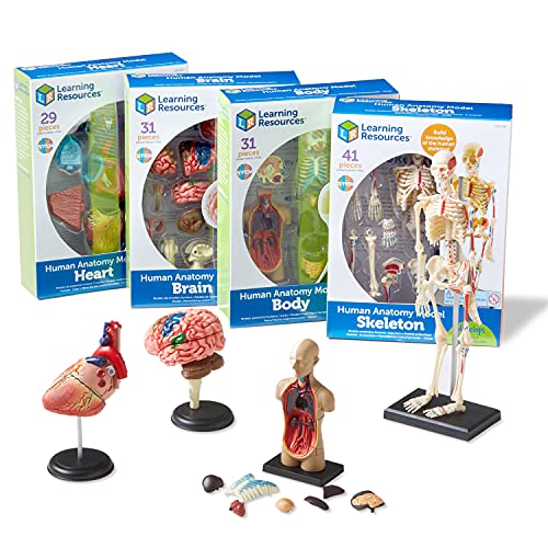
Learning Resources Anatomy Models Bundle Set – 4 STEM Anatomy Demonstration Tools, Ages 8+ Classroom Demonstration Tools, Teacher Supplies
Over the past few years, the Learning Resources Anatomy Models Bundle Set – 4 STEM Anatomy Demonstration Tools, Ages 8+ Classroom Demonstration Tools, Teacher Supplies has become one of the most popular ways for kids to learn about the human body. This miniature set measures, when assembled – Heart Model 5 inches, Brain Model 3 3/4 inches, Body Model 4 1/2 inches, Skeleton Model 92 inches. The set of four Anatomy Models is perfect for kids who want to gain a deeper understanding of how organs and systems interact by manipulating them themselves. Get ready to explore the wide world around you with science discovery toys and tools that help kids build observation and critical thinking skills used by real scientists.
Common Questions on Learning Resources Anatomy Models Bundle Set – 4 STEM Anatomy Demonstration Tools, Ages 8+ Classroom Demonstration Tools, Teacher Supplies
• What are some of the benefits of using anatomy models in the classroom?Anatomy models can provide a tangible, three-dimensional representation of concepts that can be difficult to grasp from two-dimensional textbooks. Additionally, they can be used as a starting point for discussions and debate, and can be Manipulated to demonstrate different concepts.
• What is included in this bundle set?
The Learning Resources Anatomy Models Bundle Set includes four different STEM anatomy demonstration tools, perfect for use in the classroom. The set includes a brain model, skeleton model, heart model, and lungs model.
• What age range is this bundle set appropriate for?
This bundle set is appropriate for ages 8 and up. It is a great resource for elementary and middle school classrooms.
• How can these models be used in the classroom?
These models can be used in a variety of ways in the classroom. They can be used to demonstrate and explain concepts, to start discussions and debate, and to provide a tangible, three-dimensional representation of difficult to grasp concepts.
•• What are some of the benefits of using anatomy models in the classroom?
Anatomy models can
Why We Like This
• A set of four anatomy models for kids• The models measure, when assembled: heart model 5 inches, brain model 3 3/4 inches, body model 4 1/2 inches, skeleton model 92 inches• A heart pump model and more are included to help kids gain a deeper understanding of how organs and systems interact• Learning Resources is a trusted brand by teachers and parents alike since 1984• These models are perfect for back to school and make a great addition to any science classroom
Additional Product Information
| Color | Multicolor |
| Height | 4.1 Inches |
| Length | 13.5 Inches |
| Weight | 1.6975594174 Pounds |
Anatomy and Physiology Coloring Book (Anatomy MED): Human Anatomy and Physiology Coloring Book & Workbook (Updated Edition)
The human body is an amazing machine that is made up of many different parts that work together to keep us alive and functioning. The study of anatomy and physiology is a way to learn about how these parts work together.
One way to learn about anatomy and physiology is to use a coloring book. This coloring book is filled with detailed illustrations of the human body. It also includes information on how the body works.
coloring book is a great way to learn about the human body. It is packed with detailed illustrations and information on how the body works. This coloring book is a valuable resource for anyone interested in learning about anatomy and physiology.
Common Questions on Anatomy and Physiology Coloring Book (Anatomy MED): Human Anatomy and Physiology Coloring Book & Workbook (Updated Edition)
• What are some of the benefits of using a coloring book to learn about anatomy and physiology?Coloring books can help you learn about the different parts of the body and how they work together. They can also help you memorize information and improve your understanding of anatomy and physiology.
• What should you do if you make a mistake while coloring?
If you make a mistake while coloring, you can simply erase it and try again.
Why We Like This
1. The perfect coloring book and workbook for students of human anatomy and physiology!
2. Updated edition with new illustrations and information!
3. Includes coloring pages for every major body system!
4. Perfect for use in the classroom or for studying at home!
5. Makes learning anatomy and physiology fun and engaging!
Additional Product Information
| Height | 11 inches |
| Length | 8.5 inches |
Amish Wedding Old Fashioned Bartlett Pear Halves, 32 oz. Jars (2 Jars)
Amish Wedding Old Fashioned Bartlett Pear Halves are the perfect way to enjoy the classic taste of pears. These pear halves are grown in Ohio and are hand-picked to ensure the highest quality. They are then canned in glass jars to preserve their freshness and flavor.
There are two 32oz jars included in this set, so you can enjoy the pears anytime, anywhere. With their old-fashioned taste, these pears are sure to become a family favorite.
Common Questions on Amish Wedding Old Fashioned Bartlett Pear Halves, 32 oz. Jars (2 Jars)
• What is in Amish Wedding Old Fashioned Bartlett Pear Halves, 32 oz. Jars (2 Jars)?Amish Wedding Old Fashioned Bartlett Pear Halves, 32 oz. Jars (2 Jars) contain Bartlett pears that are Halved and old-fashioned.
• How many Bartlett pears are in each jar?
There are approximately 15-20 Bartlett pears in each jar.
• What is the recipe for Amish Wedding Old Fashioned Bartlett Pear Halves?
The recipe for Amish Wedding Old Fashioned Bartlett Pear Halves is as follows: 1/2 cup sugar, 1/4 cup vinegar, 1 tablespoon grated onion, 1 tablespoon ground cloves, 1 teaspoon ground allspice, 2 (32-ounce) bottles pear halves, drained. Combine sugar, vinegar, onion, cloves, and allspice in a Dutch oven; bring to a boil. Add pear halves; return to a boil. Cover, reduce heat, and simmer 2 hours or until pears are tender. Cool slightly. Remove pears from syrup with a slotted spoon. Cover and chill pears up to 1 week. Boil syrup until
Why We Like This
Old Fashioned taste
Made in Ohio
Two 32oz Glass Jars
Perfect for canning and preserving
Great for gifting
TWOHANDS Set of 12 Micro Pens,Art Pens,Fineliner Ink Pens,Technical Drawing pen,Pigment Pen,Fine Point,Black,Waterproof,for Art Watercolor,Sketching,Anime,Manga,Scrapbooking 20413

If you’re looking for a versatile set of micro pens that can be used for everything from general writing to professional illustration, look no further than the TWOHANDS Set of 12 Micro Pens. These pens come in a variety of sizes, from 02mm to 30mm, so you can find the perfect one for whatever task you’re working on. Plus, each pen is made with archival quality ink that is waterproof and fade resistant, so your work will always look its best. The set also comes with a handy storage pouch, making it easy to keep all your pens organized and ready to go.
Common Questions on TWOHANDS Set of 12 Micro Pens,Art Pens,Fineliner Ink Pens,Technical Drawing pen,Pigment Pen,Fine Point,Black,Waterproof,for Art Watercolor,Sketching,Anime,Manga,Scrapbooking 20413
• What is the name of the company that makes TWOHANDS Set of 12 Micro Pens,Art Pens,Fineliner Ink Pens,Technical Drawing pen,Pigment Pen,Fine Point,Black,Waterproof,for Art Watercolor,Sketching,Anime,Manga,Scrapbooking 20413?The company’s name isTWOHANDS.
• Where are TWOHANDS Set of 12 Micro Pens,Art Pens,Fineliner Ink Pens,Technical Drawing pen,Pigment Pen,Fine Point,Black,Waterproof,for Art Watercolor,Sketching,Anime,Manga,Scrapbooking 20413 made?
TWOHANDS Set of 12 Micro Pens,Art Pens,Fineliner Ink Pens,Technical Drawing pen,Pigment Pen,Fine Point,Black,Waterproof,for Art Watercolor,Sketching,Anime,Manga,Scrapbooking 20413 are made in China.
• What is the warranty period for TWOHANDS Set of 12 Micro Pens,Art Pens,Fineliner Ink Pens,Technical Drawing pen,Pigment Pen,Fine Point,Black,Waterproof,
Why We Like This
• 1. Fine point black pigment ink pens• 2. Perfect for bullet journals, general writing, and technical drawing• 3. Archival quality ink is waterproof and fade resistant• 4. Each set comes in a handy storage pouch• 5. Makes a great gift for any occasion
Additional Product Information
| Color | Black |
Benefits of Radiographic Positioning And Related Anatomy
Radiographic positioning and related anatomy play an important role in providing quality images for diagnosis. Proper patient positioning ensures that the desired body part is correctly positioned in the x-ray beam, while also minimizing radiation exposure to other parts of the body. Additionally, knowledge of related anatomy helps radiographers to optimize image acquisition by understanding how different structures will appear on a radiograph.
Some benefits of proper radiographic positioning and related anatomy include:
• Improved diagnostic accuracy – By ensuring that the correct body part is correctly positioned in the x-ray beam, radiographers can minimize artifacts and improve diagnostic accuracy.
• Enhanced image quality – Knowledge of related anatomy can help radiographers optimize image acquisition, resulting in clearer images that are easier to interpret.
Buying Guide for Best Radiographic Positioning And Related Anatomy
It is important to have a basic understanding of radiographic positioning and related anatomy in order to produce high-quality images. The following are some tips on how to improve your radiographic positioning:
1. Use a lead shield when performing x-rays on pregnant patients or young children.
2. Make sure the x-ray beam is perpendicular to the body part being imaged.
3. Keep the x-ray film as close to the body as possible.
4. Use appropriate collimation techniques to avoid unnecessary radiation exposure.
5. Be sure to identify all anatomic structures on the x-ray film before making any adjustments.
Frequently Asked Question
Which radiographic positioning technique will best depict the desired anatomy?
The radiographic positioning technique that will best depict the desired anatomy will depend on the specific anatomy that is being imaged. For example, if the anatomy is located in the chest, then a chest x-ray may be the best radiographic positioning technique.
What are the potential pitfalls of each radiographic technique?
There are several potential pitfalls for each radiographic technique. One is that the technique may not be able to capture all of the desired information. Another is that the technique may be less effective at lower doses of radiation. Finally, the technique may be more likely to cause artifacts or false positives.
How can you optimize the image quality for each radiographic technique?
Some tips on how to optimize image quality for radiographic techniques include: – using the highest quality imaging equipment possible- using proper technique when performing the radiographic examination- ensuring that the patient is well positioned- using appropriate exposure settings- using appropriate filtration
What is the typical radiation dose for each radiographic technique?
In general, the radiation dose for a typical radiographic procedure is relatively low and is not generally considered to be a cause for concern.
Are there any special considerations for pregnant patients or children when performing radiographic imaging?
There are special considerations for pregnant patients or children when performing radiographic imaging. Pregnant patients should avoid x-rays during pregnancy, as they can be harmful to the developing fetus. Children should also avoid x-rays, as their bodies are still developing and are more sensitive to the effects of radiation.
Conclusion
We hope that you have enjoyed learning about the best radiographic positioning and related anatomy and found our products to be of interest. We would be happy to answer any questions that you may have or provide you with additional information.
Thank you for your time, we look forward to hearing from you soon.